Neuroscience - Cell-Based Imaging Methods for Investigating the Structure and Function of Neuronal Cells
Over the last decade, advances in imaging and labeling techniques have made it possible to resolve structures and processes with high spatio-temporal resolution, even in living cells. In this article, we summarize microscopy-related methods, and cite recent research findings that have helped researchers gain a deeper understanding of the complex organization and function of the mammalian nervous system.

Rat fibroblast, surrounded by parallel-aligned rat Schwann cells (SCs), cultured in an ibidi µ-Slide 8 Well and stained for the SC-marker S100 (green), Vimentin (grey), and DAPI (blue). The image was obtained with a laser-scanning microscope. Courtesy: Flavia Millesi, Medical University Vienna, Austria
Visualization of Subcellular Structures and Components
Fluorescent Labeling
Fluorescent labeling is a commonly used, easy to perform method for revealing subcellular structures and components, and their interaction partners, in mostly fixed cells or tissue. The labels include single particles (e.g., quantum dots or dye-labeled origamis), organic dyes (e.g., Rhodamine or Alexa dyes), and fluorescently labeled antibodies. For example, Bhat et al. visualized actin structures of neuronal CAD (Cath.a-differentiated) cells with Rhodamine Phalloidin, which allowed for the detection and quantification of tunneling nanotubes connected cells (TNT)1.
In order to visualize and characterize multiple cellular components in the same biological sample simultaneously, an immunolabeling approach using different primary and secondary antibodies is often employed. For example, Millesi et al. developed multi-color staining panels by expressing specific markers to discriminate between Schwann Cells, fibroblasts, and dorsal root ganglion neurons in the same cell culture2. Immunostaining can also be performed on whole tissues (immunohistology). In order to quantify brainstem neurons, Usseglio et al. dissected the brains of adult mice, then fixed, cryo-embedded, and sectioned them prior to immunostaining3.
Read more about immunofluorescence here.
Endogenous Labeling of Living Cells
For live-cell imaging experiments, in particular, a labeling approach that does not interfere with the viability of the cells and their functions is required. Small-molecule binders (e.g., SiR-actin or LifeAct) coupled to a fluorescent protein (e.g., GFP or RFP) can be used for this approach. LifeAct stains filamentous actin (F-actin) structures in living (or fixed) eukaryotic cells and tissues. For example, LifeAct tagged with RFP was used to visualize the F-actin cytoskeleton for dendritic spine analysis in living primary hippocampal neurons4.
Read more about actin visualization in living cells here.
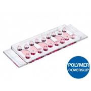
Imaging of 3D Neural Co-Cultures
When mimicking the complex microenvironment of living tissue, it is essential to investigate the cells of interest, plus any other cells they interact with in the surrounding cellular matrix. Therefore, generating 3D models of the co-cultures of these interacting cells within a physiological scaffold is an important strategy for understanding cellular structure and function.
To study the interaction of human peripheral sensory neurons (PSNs) and endothelial cells (ECs), and to investigate whether PSNs affect angiogenesis, Kannan et al. developed a 3D co-culture system that is comprised of human embryonic stem cell (hESC) PSNs and hESC endothelial cells embedded in a collagen type 1 matrix5.
Millesi et al. performed live-cell imaging and manual tracking of a fibroblast-Schwann cell co-culture to investigate if rat fibroblasts affect the migratory behavior of rat Schwann cells cultivated on regenerative spider silk2.
Read more about 3D cell culture here.
Analyzing Neuronal Cell Function and Organization Using High Spatiotemporal Resolution
Live Cell Imaging
Live cell imaging of neuronal cells and tissues helps to provide mechanistic insights into neuronal processes and functions. For example, fluorescence lifetime imaging microscopy (FLIM) in brain slices revealed metabolic changes were the cause of mitochondrial dysfunction in neurodegenerative diseases6.
Physiological Conditions
For long-term live cell imaging, specifically, it is crucial to maintain physiological conditions by controlling the temperature, pH, oxygen, and humidity. For example, the live cell imaging of central nervous system (CNS) neurons with stable environmental conditions of 37°C and 5% oxygen allowed for the visualization of axon polarization and extension in 3D over a time-course of 48h7. CNS neurons were embedded in collagen hydrogels, then imaged with DIC under physiological conditions to show that axon growth behavior in 3D is amoeboid and independent of adhesions7.
Read more about live cell imaging and physiological conditions on the microscope here.
In order to simulate physiological conditions for endothelial cells, such as Brain Microvascular Endothelial Cells (BMECs) in vitro, it is crucial to cultivate them under defined shear stress conditions with continuous media flow. For example, Gao et al. showed that altered cerebral blood flow in gerbil brains after global cerebral IR injuries affects microvascular endothelial and neuronal function8.
Read more about cell culture under flow here.
Super-Resolution Imaging of Neuronal Cells
In the nervous system, many neuronal structures and processes are smaller than the resolution limit of light. Therefore, it is difficult to resolve details that are smaller, or closer, than 200nm together with conventional light microscopy techniques. The development of super-resolution methods, such as structured illumination microscopy (SIM), stimulated emission depletion (STED) microscopy, or photoactivated localization microscopy (PALM), have made it possible to overcome the diffraction barrier of the light, and therefore made it possible to visualize components as small as synaptic vesicles with a size of 40nm. For example, Conaghi et al. were able to localize individual SENP proteins in cultured hippocampal primary neurons with structured illumination microscopy (SIM) for the first time9.
Read more about super-resolution imaging here.
Calcium Imaging in Neurons
Calcium signaling plays a key role in almost all cellular tasks. In neurons, for example, Ca2+ ions are responsible for the release of neurotransmitters from synaptic vesicles.
Calcium imaging enables the monitoring of the electrical activity of neurons by following Ca2+ flux with fluorescent probes that are activated upon Ca2+ binding. Calcium imaging can be used as a tool to evaluate cell functionality, for example, when differentiating human induced pluripotent stem cells (iPSCs) into neurons10.
Find out more about ibidi solutions for Neurobiological research here.
Cited References:
- B. Bhat, N. Ljubojevic, S. Zhu, M. Fukuda, A. Echard, C. Zurzolo. Rab35 and its effectors promote formation of tunneling nanotubes in neuronal cells. Scientific reports, 2020, 10.1038/s41598-020-74013-z Read article
- F . Millesi, T . Weiss, A. Mann, M. Haertinger, L. Semmler, P. Supper, D.Pils, A. Naghilou, C. Radtke. Defining the regenerative effects of native spider silk fibers on primary Schwann cells, sensory neurons, and nerve-associated fibroblasts. FASEB Journal. 2021, 10.1096/fj.202001447R Read article
- G. Usseglio, E. Gatier, A. Heuzé, C. Hérent, J. Bouvier. Control of Orienting Movements and Locomotion by Projection-Defined Subsets of Brainstem V2a Neurons. Current Biology, 2020. 10.1016/j.cub.2020.09.014. Read article
- D. Reim, T.M. Weis, S. Halbedl, J.P. Delling, A.M. Grabrucker, T.M. Boeckers, M.J. Schmeisser. The Shank3 Interaction Partner ProSAPiP1 Regulates Postsynaptic S.PAR Levels and the Maturation of Dendritic Spines in Hippocampal Neurons. Frontiers in Synaptic Neuroscice, 2016. 10.3389/fnsyn.2016.00013 Read article
- S. Kannan, M. Lee, S. Muthusamy, A. Blasiak, G. Sriram, T. Cao. Peripheral sensory neurons promote angiogenesis in neurovascular models derived from hESCs. Stem Cell Research, 2021, 10.1016/j.scr.2021.102231. Read article
- E. Motori, I. Atanassov, S. M. V. Kochan, K. Folz-Donahue, V. Sakthivelu, P. Giavalisco, N. Toni, J. Puyal, N.-G. Larsson. Neuronal metabolic rewiring promotes resilience to neurodegeneration caused by mitochondrial dysfunction. Science Advances, 2020, 10.1126/sciadv.aba8271 Read article
- T. E. Santos, B. Schaffran, N. Broguière, L. Meyn, M. Zenobi-Wong, F. Bradke. Axon Growth of CNS Neurons in Three Dimensions Is Amoeboid and Independent of Adhesions. Cell Reports, 2021, 10.1016/j.celrep.2020.107907 Read article
- J. Q. Gao, P. Wang, J. W. Yan, L. N. Ba, P. L. Shi, H. M. Wu, X. Y. Guan, Y. G. Cao, H.L. Sun, X. Y. Mao. Shear Stress Rescued the Neuronal Impairment Induced by Global Cerebral Ischemia Reperfusion via Activating PECAM-1-eNOS-NO Pathway. Frontiers in cell and developmental biology, 2021, 10.3389/fcell.2020.631286 Read article
- L. Colnaghi, A. Conz, L. Russo, C.A. Musi, L. Fioriti, T. Borsello, M. Salmona. Neuronal Localization of SENP Proteins with Super Resolution Microscopy. Brain Sciences, 2020, 10.3390/brainsci10110778 Read article
- F. Bianchi, M. Malboubi, Y. Li, J. H. George, A. Jerusalem, F. Szele, M. S. Thompson, H. Ye. Rapid and efficient differentiation of functional motor neurons from human iPSC for neural injury modelling. Stem Cell Research, 2018, 10.1016/j.scr.2018.09.006 Read article
Original article you will find on ibidi website.